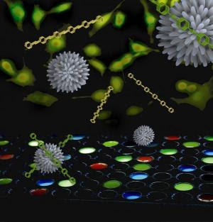
Using a “chemical nose” array of nanoparticles and polymers, researchers at the University of Massachusetts Amherst have developed a fundamentally new, more effective way to differentiate not only between healthy and cancerous cells but also between metastatic and non-metastatic cancer cells. It’s a tool that could revolutionize cancer detection and treatment, according to chemist Vincent Rotello and cancer specialist Joseph Jerry.An article describing Rotello and colleagues’ new chemical nose method of cancer detection appears in the June 23 issue of the journal Proceedings of the National Academy of Sciences online.
Currently, detecting cancer via cell surface biomarkers has taken what’s known as the “lock and key” approach. Drawbacks of this method include that foreknowledge of the biomarker is required. Also, as Rotello explains, a cancer cell has the same biomarkers on its surface as a healthy cell, but in different concentrations, a maddeningly small difference that can be very difficult to detect. “You often don’t get a big signal for the presence of cancer,” he notes. “It’s a subtle thing.”
He adds, “Our new method uses an array of sensors to recognize not only known cancer types, but it signals that abnormal cells are present. That is, the chemical nose can simply tell us something isn’t right, like a ‘check engine light,’ though it may never have encountered that type before.” Further, the chemical nose can be designed to alert doctors of the most invasive cancer types, those for which early treatment is crucial.
In blinded experiments in four human cancer cell lines (cervical, liver, testis and breast), as well as in three metastatic breast cell lines, and in normal cells, the new detection technique correctly indicated not only the presence of cancer cells in a sample but also identified primary cancer vs. metastatic disease.
In further experiments to rule out the possibility that the chemical nose had simply detected individual differences in cells from different donors, the researchers repeated the experiments in skin cells from three groups of cloned BALB /c mice: healthy animals, those with primary cancer and those with metastatic disease. Once again, it worked. “This result is key,” says Rotello. “It shows that we can differentiate between the the three cell types in a single individual using the chemical nose approach.”
Rotello’s research team, with colleagues at the Georgia Institute of Technology, designed the new detection system by combining three gold nanoparticles that have special affinity for the surface of chemically abnormal cells, plus a polymer known as PPE, or para-phenyleneethynylene. As the ‘check engine light,’ PPE fluoresces or glows when displaced from the nanoparticle surface.
By adding PPE bound with gold nanoparticles to human cells incubating in wells on a culture plate, the researchers induce a response called “competitive binding.” Cell surfaces bind the nanoparticles, displacing the PPE from the surface. This turns on PPE’s fluorescent switch. Cells are then identified from the patterns generated by different particle-PPE systems.
Rotello says the chemical nose approach is so named because it works like a human nose, which is arrayed with hundreds of very selective chemical receptors. These bind with thousands of different chemicals in the air, some more strongly than others, in the endless combination we encounter. The receptors report instantly to the brain, which recognizes patterns such as, for example, “French fries,” or it creates a new smell pattern.
Chemical receptors in the nose plus the brain’s pattern recognition skills together are incredibly sensitive at detecting subtly different combinations, Rotello notes. We routinely detect the presence of tiny numbers of bacteria in meat that’s going bad, for instance. Like a human nose, the chemical version being developed for use in cancer also remembers patterns experienced, even if only once, and creates a new one when needed.
For the future, Rotello says further studies will be undertaken in an animal model to see if the chemical nose approach can identify cell status in real tissue. Also, more work is required to learn how to train the chemical nose’s sensors to give more precise information to physicians who will be making judgment calls about patients’ cancer treatment. But the future is promising, he adds. “We’re getting complete identification now, and this can be improved by adding more and different nanoparticles. So far we’ve experimented with only three, and there are hundreds more we can make.”
INTERNAL MEDICINE The internal medicine blog , where you can have details on alternatice medicine , latest trends in medicine , new drugs in the market , school of medicine etc
‘Chemical Nose’ May Sniff Out Cancer Earlier
Wednesday, June 24, 2009 at 12:58 AM Posted by Sajith
Autonomous Robot Detects Shrapnel In Flesh
at 12:54 AM Posted by Sajith

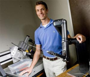
Bioengineers at Duke University have developed a laboratory robot that can successfully locate tiny pieces of metal within flesh and guide a needle to its exact location -– all without the need for human assistance.The successful proof-of-feasibility experiments lead the researchers to believe that in the future, such a robot could not only help treat shrapnel injuries on the battlefield, but might also be used for such medical procedures as placing and removing radioactive "seeds" used in the treatment of prostate and other cancers.
In their latest experiments, the engineers started with a rudimentary tabletop robot whose "eyes" are a novel 3-D ultrasound technology developed at Duke. An artificial intelligence program served as the robot's "brain" by taking the real-time 3-D information, processing it and giving the robot specific commands to perform. In their simulations, the researchers used tiny (2 millimeter) pieces of needle because, like shrapnel, they are subject to magnetism.
"We attached an electromagnet to our 3-D probe, which caused the shrapnel to vibrate just enough that its motion could be detected," said A.J. Rogers, who just completed an undergraduate degree in bioengineering at Duke. "Once the shrapnel's coordinates were established by the computer, it successfully guided a needle to the site of the shrapnel."
By proving that the robot could guide a needle to an exact location, it would simply be a matter of replacing the needle probe with a tiny tool, such as a grabber, the researchers said.
Rogers worked in the laboratory of Stephen Smith, director of the Duke University Ultrasound Transducer Group and senior member of the research team. The results of the experiments were published early online in the July issue of the journal IEEE Transactions on Ultrasonics, Ferroelectrics and Frequency Control.
Since the researchers achieved positive results using a rudimentary robot and a basic artificial intelligence program, they are encouraged that simple and reasonably safe procedures will become routine in the near future as robot and artificial intelligence technology improves.
"We showed that in principle, the system works," Smith said. "It can be very difficult using conventional means to detect small pieces of shrapnel, especially in the field. The military has an extensive program of exploring the use of surgical robots in the field, and this advance could play a role."
In addition to its applications recovering the radioactive seeds used in treating prostate cancer, Smith said the system could also prove useful in removing foreign, metallic objects from the eye.
Advances in ultrasound technology have made these latest experiments possible, the researchers said, by generating detailed, 3-D moving images in real-time. The Duke team has a long track record of modifying traditional 2-D ultrasound – like that used to image babies in utero – into the more advanced 3-D scans. Since inventing the technique in 1991, the team has shown its utility by developing specialized catheters and endoscopes for real-time imaging of blood vessels in the heart and brain.
In the latest experiments, the robot successfully performed its main task: locating a tiny piece of metal in a water bath, then directing a needle on the end of the robotic arm to it. The researchers had previously used this approach to detect micro-calcifications in simulated breast tissue. In the latest experiments, Rogers added an electromagnet to the end of the transducer, or wand, the device that sends out and receives the ultrasonic waves.
"The movement caused by the electromagnet on the shrapnel was not visible to the human eye," Rogers said. "However, on the 3-D color Doppler system, the moving shrapnel stood out plainly as bright red."
The robot used in these experiments is a tabletop version capable of moving in three axes. For the next series of tests, the Duke researchers plan to use a robotic arm with six-axis capability.
The research in Smith's lab is supported by the National Institutes of Health. Duke's Ned Light was also part of the research team.
Search
Labels
- Alzheimer's (10)
- Antibiotics (7)
- Anxiety (1)
- articles (2)
- Bacteria (1)
- behavioral abnormality (1)
- Bio technology (1)
- Brain (8)
- breast cancer (6)
- Cancer (40)
- cancer treatment (7)
- chemotherapy (6)
- Chlamydia (1)
- Cytology (3)
- Death (1)
- Diabetes Mellitus (7)
- Diet (17)
- DNA (1)
- Doctors (1)
- epilepsy (2)
- Fossil (1)
- fruits (1)
- genes (6)
- Genetics (13)
- Geriatrics (1)
- Health (12)
- Heart diseases (9)
- Herbal Medicine (2)
- HIV and AIDS (7)
- influenza (1)
- Inventions (1)
- IVF (1)
- latest findings (1)
- Life style (2)
- Lung Cancer (1)
- Lung Disease (9)
- Microbiology (1)
- Multiple Sclerosis (1)
- Nanotechnology (3)
- Neonatology (1)
- neurology (1)
- News (18)
- Parkinsonism (2)
- Prevention (1)
- Primates (1)
- Prostate cancer (1)
- Prosthetics (1)
- seizure (2)
- Skin (1)
- STD (2)
- Stem cells (9)
- Stroke (1)
- Surgery (2)
- Swine flu (11)
- Virus (1)
- weight loss (1)
Search The Web
Blog Archive
Minyx v2.0 template es un theme creado por Spiga. | Minyx Blogger Template distributed by eBlog Templates
