Spiga
Showing posts with label Nanotechnology. Show all posts

Nanoparticles Shrink Tumors in Mice

Share

The application of nanotechnology in the field of drug delivery has attracted much attention in recent years. In cancer research, nanotechnology holds great promise for the development of targeted, localized delivery of anticancer drugs, in which only cancer cells are affected.

A dorsal view of a mouse showing accumulation of nanoparticles in a tumor four hours after intravenous administration. Bright fluorescence is observed predominantly in the tumor.

Such targeted-therapy methods would represent a major advance over current chemotherapy, in which anticancer drugs are distributed throughout the body, attacking healthy cells along with cancer cells and causing a number of adverse side effects.
By carrying out comprehensive studies on mice with human tumors, UCLA scientists have obtained results that move the research one step closer to this goal. In a paper published July 8 in the journal Small, researchers at UCLA's California NanoSystems Institute and Jonsson Comprehensive Cancer Center demonstrate that mesoporous silica nanoparticles (MSNs), tiny particles with thousands of pores, can store and deliver chemotherapeutic drugs in vivo and effectively suppress tumors in mice.
The researchers also showed that MSNs accumulate almost exclusively in tumors after administration and that the nanoparticles are excreted from the body after they have delivered their chemotherapeutic drugs.
The study was conducted jointly in the laboratories of Fuyu Tamanoi, a UCLA professor of microbiology, immunology and molecular genetics and director of the signal transduction and therapeutics program at UCLA's Jonsson Comprehensive Cancer Center, and Jeffrey Zink, a UCLA professor of chemistry and biochemistry. Tamanoi and Zink are researchers at the California NanoSystems Institute (CNSI) and are two of the co-directors of the CNSI's Nano Machine Center for Targeted Delivery and On-Demand Release. The lead investigator on the research is Jie Lu, a postdoctoral fellow in Tamanoi's lab. Monty Liong and Zongxi Li, researchers from Zink's lab, also contributed to this work.
In the study, researchers found that MSNs circulate in the bloodstream for extended periods of time and accumulate predominantly in tumors. The tumor accumulation could be further improved by attaching a targeting moiety to MSNs, the researchers said.
The treatment of mice with camptothecin-loaded MSNs led to shrinkage and regression of xenograft tumors. By the end of the treatment, the mice were essentially tumor free, and acute and long-term toxicity of MSNs to the mice was negligible. Mice with breast cancer were used in this study, but the researchers have recently obtained similar results using mice with human pancreatic cancer.
"Our present study shows, for the first time, that MSNs are effective for anticancer drug delivery and that the capacity for tumor suppression is significant," Tamanoi said.
"Two properties of these nanoparticles are important," Lu said. "First, their ability to accumulate in tumors is excellent. They appear to evade the surveillance mechanism that normally removes materials foreign to the body. Second, most of the nanoparticles that were injected into the mice were excreted out through urine and feces within four days. The latter results are quite interesting and might explain the low toxicity observed in the biocompatabilty experiments we conducted."
Researchers at the Nano Machine Center for Targeted Delivery and On-Demand Release are modifying MSNs -- which are easily modifiable -- so that the nanoparticles can be equipped with nanomachines. For example, nanovalves are being attached at the opening of the pores to control the release of anticancer drugs. In addition, the interior of the pores is being modified so that the light-induced release of anticancer drugs can be achieved.
"We can modify both the particles themselves and also the attachments on the particles in a wide variety of ways, which makes this material particularly attractive for engineering drug-delivery vehicles," Zink said.
The team is now planning future research that involves testing MSNs in a variety of animal-model systems and carrying out extensive studies on the safety of MSNs.
"Comprehensive investigation with practical dosages which are adequate and suitable for in vivo delivery of anticancer drugs is needed before MSNs can reach clinics as a drug-delivery system," Tamanoi said.
The research received support from National Institutes of Health and the National Science Foundation. In addition, NanoPacific Holdings Inc. provided critical support for the animal experiments.
‘Chemical Nose’ May Sniff Out Cancer Earlier

Share

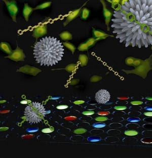
Nanoparticles and polymers were used to create a sensor that can distinguish between healthy, cancerous and metastatic cells.
Using a “chemical nose” array of nanoparticles and polymers, researchers at the University of Massachusetts Amherst have developed a fundamentally new, more effective way to differentiate not only between healthy and cancerous cells but also between metastatic and non-metastatic cancer cells. It’s a tool that could revolutionize cancer detection and treatment, according to chemist Vincent Rotello and cancer specialist Joseph Jerry.An article describing Rotello and colleagues’ new chemical nose method of cancer detection appears in the June 23 issue of the journal Proceedings of the National Academy of Sciences online.

Currently, detecting cancer via cell surface biomarkers has taken what’s known as the “lock and key” approach. Drawbacks of this method include that foreknowledge of the biomarker is required. Also, as Rotello explains, a cancer cell has the same biomarkers on its surface as a healthy cell, but in different concentrations, a maddeningly small difference that can be very difficult to detect. “You often don’t get a big signal for the presence of cancer,” he notes. “It’s a subtle thing.”

He adds, “Our new method uses an array of sensors to recognize not only known cancer types, but it signals that abnormal cells are present. That is, the chemical nose can simply tell us something isn’t right, like a ‘check engine light,’ though it may never have encountered that type before.” Further, the chemical nose can be designed to alert doctors of the most invasive cancer types, those for which early treatment is crucial.

In blinded experiments in four human cancer cell lines (cervical, liver, testis and breast), as well as in three metastatic breast cell lines, and in normal cells, the new detection technique correctly indicated not only the presence of cancer cells in a sample but also identified primary cancer vs. metastatic disease.

In further experiments to rule out the possibility that the chemical nose had simply detected individual differences in cells from different donors, the researchers repeated the experiments in skin cells from three groups of cloned BALB /c mice: healthy animals, those with primary cancer and those with metastatic disease. Once again, it worked. “This result is key,” says Rotello. “It shows that we can differentiate between the the three cell types in a single individual using the chemical nose approach.”

Rotello’s research team, with colleagues at the Georgia Institute of Technology, designed the new detection system by combining three gold nanoparticles that have special affinity for the surface of chemically abnormal cells, plus a polymer known as PPE, or para-phenyleneethynylene. As the ‘check engine light,’ PPE fluoresces or glows when displaced from the nanoparticle surface.

By adding PPE bound with gold nanoparticles to human cells incubating in wells on a culture plate, the researchers induce a response called “competitive binding.” Cell surfaces bind the nanoparticles, displacing the PPE from the surface. This turns on PPE’s fluorescent switch. Cells are then identified from the patterns generated by different particle-PPE systems.

Rotello says the chemical nose approach is so named because it works like a human nose, which is arrayed with hundreds of very selective chemical receptors. These bind with thousands of different chemicals in the air, some more strongly than others, in the endless combination we encounter. The receptors report instantly to the brain, which recognizes patterns such as, for example, “French fries,” or it creates a new smell pattern.

Chemical receptors in the nose plus the brain’s pattern recognition skills together are incredibly sensitive at detecting subtly different combinations, Rotello notes. We routinely detect the presence of tiny numbers of bacteria in meat that’s going bad, for instance. Like a human nose, the chemical version being developed for use in cancer also remembers patterns experienced, even if only once, and creates a new one when needed.

For the future, Rotello says further studies will be undertaken in an animal model to see if the chemical nose approach can identify cell status in real tissue. Also, more work is required to learn how to train the chemical nose’s sensors to give more precise information to physicians who will be making judgment calls about patients’ cancer treatment. But the future is promising, he adds. “We’re getting complete identification now, and this can be improved by adding more and different nanoparticles. So far we’ve experimented with only three, and there are hundreds more we can make.”

New Nanoparticles Could Lead To End Of Chemotherapy

Share

Dr. Manuel Perez and his team
Nanoparticles specially engineered by University of Central Florida Assistant Professor J. Manuel Perez and his colleagues could someday target and destroy tumors, sparing patients from toxic, whole-body chemotherapies.Perez and his team used a drug called Taxol for their cell culture studies, recently published in the journal Small, because it is one of the most widely used chemotherapeutic drugs. Taxol normally causes many negative side effects because it travels throughout the body and damages healthy tissue as well as cancer cells.

The Taxol-carrying nanoparticles engineered in Perez's laboratory are modified so they carry the drug only to the cancer cells, allowing targeted cancer treatment without harming healthy cells. This is achieved by attaching a vitamin (folic acid) derivative that cancer cells like to consume in high amounts.

Because the nanoparticles also carry a fluorescent dye and an iron oxide magnetic core, their locations within the cells and the body can be seen by optical imaging and magnetic resonance imaging (MRI). That allows a physician to see how the tumor is responding to the treatment.

The nanoparticles also can be engineered without the drug and used as imaging (contrast) agents for cancer. If there is no cancer, the biodegradable nanoparticles will not bind to the tissue and will be eliminated by the liver. The iron oxide core will be utilized as regular iron in the body.

"What's unique about our work is that the nanoparticle has a dual role, as a diagnostic and therapeutic agent in a biodegradable and biocompatible vehicle," Perez said.

Perez has spent the past five years looking at ways nanotechnology can be used to help diagnose, image and treat cancer and infectious diseases. It's part of the quickly evolving world of nanomedicine.

The process works like this. Cancer cells in the tumor connect with the engineered nanoparticles via cell receptors that can be regarded as "doors" or "docking stations." The nanoparticles enter the cell and release their cargo of iron oxide, fluorescent dye and drugs, allowing dual imaging and treatment.

"Although the results from the cell cultures are preliminary, they are very encouraging," Perez said.

A new chemistry called "click chemistry" was utilized to attach the targeting molecule (folic acid) to the nanoparticles. This chemistry allows for the easy and specific attachment of molecules to nanoparticles without unwanted side products. It also allows for the easy attachment of other molecules to nanoparticles to specifically seek out particular tumors and other malignancies.

Perez's study builds on his prior research published in the prestigious journal Angewandte Chemie Int. Ed. His work has been partially funded by a National Institutes of Health grant and a Nanoscience Technology Center start-up fund.

"Our work is an important beginning, because it demonstrates an avenue for using nanotechnology not only to diagnose but also to treat cancer, potentially at an early stage," Perez said.

Perez, a Puerto Rico native, joined UCF in 2005. He works at UCF's NanoScience Technology Center and Chemistry Department and in the Burnett School of Biomedical Sciences in the College of Medicine. He has a Ph.D. from Boston University in Biochemistry and completed postdoctoral training at Massachusetts General Hospital, Harvard Medical School's teaching and research hospital.