Spiga
Showing posts with label Genetics. Show all posts

Primates' Unique Gene Regulation Mechanism

Share

Three baby orangutans from Tanjung Putting Orangutan Rehab Center in Borneo Indonesia. Scientists have discovered a new way genes are regulated that is unique to primates, including humans and monkeys. 
Scientists have discovered a new way genes are regulated that is unique to primates, including humans and monkeys. Though the human genome -- all the genes that an individual possesses -- was sequenced 10 years ago, greater understanding of how genes function and are regulated is needed to make advances in medicine, including changing the way we diagnose, treat and prevent a wide range of diseases.
"It's extremely valuable that we've sequenced a large bulk of the human genome, but sequence without function doesn't get us very far, which is why our finding is so important," said Lynne E. Maquat, Ph.D., lead author of the new study published February 9 in the journal Nature.
When our genes go awry, many diseases, such as cancer, Alzheimer's and cystic fibrosis can result. The study introduces a unique regulatory mechanism that could prove to be a valuable treatment target as researchers seek to manipulate gene expression -- the conversion of genetic information into proteins that make up the body and perform most life functions -- to improve human health.
The newly identified mechanism involves Alu elements, repetitive DNA elements that spread throughout the genome as primates evolved. While scientists have known about the existence of Alu elements for many years, their function, if any, was largely unknown.
Maquat discovered that Alu elements team up with molecules called long noncoding RNAs (lncRNAs) to regulate protein production. They do this by ensuring messenger RNAs (mRNAs), which take genetic instructions from DNA and use it to create proteins, stay on track and create the right number of proteins. If left unchecked, protein production can spiral out of control, leading to the proliferation or multiplication of cells, which is characteristic of diseases such as cancer.
"Previously, no one knew what Alu elements and long noncoding RNAs did, whether they were junk or if they had any purpose. Now, we've shown that they actually have important roles in regulating protein production," said Maquat, the J. Lowell Orbison Chair, professor of Biochemistry and Biophysics and director of the Center for RNA Biology at the University of Rochester Medical Center.
The expression of genes that call for the development of proteins involves numerous steps, all of which are required to occur in a precise order to achieve the appropriate timing and amount of protein production. Each of these steps is regulated, and the pathway discovered is one of only a few pathways known to regulate mRNAs directly in the midst of the protein production process.
Regulating mRNAs is one of several ways cells control gene expression, and researchers from institutions and companies around the world are honing in on this regulatory landscape in search of new ways to manage and treat disease.
According to Maquat, "This new mechanism is really a surprise. We continue to be amazed by all the different ways mRNAs can be regulated."
Maquat and the study's first author, Chenguang Gong, a graduate student in the Department of Biochemistry and Biophysics at the Medical Center, found that long noncoding RNAs and Alu elements work together to trigger a process known as SMD (Staufen 1-mediated mRNA decay). SMD conditionally destroys mRNAs after they orchestrate the production of a certain amount of proteins, preventing the creation of excessive, unwanted proteins in the body that can disrupt normal processes and initiate disease.
Specifically, long noncoding RNAs and Alu elements recruit the protein Staufen-1 to bind to numerous mRNAs. Once an mRNA finishes directing a round of protein production, Staufen-1 works with another regulatory protein previously identified by Maquat, UPF1, to initiate the degradation or decay of the mRNA so that it cannot create any more proteins.
While the research fills in a piece of the puzzle as to how our genes operate, it also accentuates the overwhelming complexity of how our DNA shapes us and the many known and unknown players involved. Maquat and Gong plan on exploring the newly identified pathway in future research.
This research was supported by a grant from the General Medical Sciences Division of the National Institutes of Health and an Elon Huntington Hooker Graduate Student Fellowship.

Nanoparticles Shrink Tumors in Mice

Share

The application of nanotechnology in the field of drug delivery has attracted much attention in recent years. In cancer research, nanotechnology holds great promise for the development of targeted, localized delivery of anticancer drugs, in which only cancer cells are affected.

A dorsal view of a mouse showing accumulation of nanoparticles in a tumor four hours after intravenous administration. Bright fluorescence is observed predominantly in the tumor.

Such targeted-therapy methods would represent a major advance over current chemotherapy, in which anticancer drugs are distributed throughout the body, attacking healthy cells along with cancer cells and causing a number of adverse side effects.
By carrying out comprehensive studies on mice with human tumors, UCLA scientists have obtained results that move the research one step closer to this goal. In a paper published July 8 in the journal Small, researchers at UCLA's California NanoSystems Institute and Jonsson Comprehensive Cancer Center demonstrate that mesoporous silica nanoparticles (MSNs), tiny particles with thousands of pores, can store and deliver chemotherapeutic drugs in vivo and effectively suppress tumors in mice.
The researchers also showed that MSNs accumulate almost exclusively in tumors after administration and that the nanoparticles are excreted from the body after they have delivered their chemotherapeutic drugs.
The study was conducted jointly in the laboratories of Fuyu Tamanoi, a UCLA professor of microbiology, immunology and molecular genetics and director of the signal transduction and therapeutics program at UCLA's Jonsson Comprehensive Cancer Center, and Jeffrey Zink, a UCLA professor of chemistry and biochemistry. Tamanoi and Zink are researchers at the California NanoSystems Institute (CNSI) and are two of the co-directors of the CNSI's Nano Machine Center for Targeted Delivery and On-Demand Release. The lead investigator on the research is Jie Lu, a postdoctoral fellow in Tamanoi's lab. Monty Liong and Zongxi Li, researchers from Zink's lab, also contributed to this work.
In the study, researchers found that MSNs circulate in the bloodstream for extended periods of time and accumulate predominantly in tumors. The tumor accumulation could be further improved by attaching a targeting moiety to MSNs, the researchers said.
The treatment of mice with camptothecin-loaded MSNs led to shrinkage and regression of xenograft tumors. By the end of the treatment, the mice were essentially tumor free, and acute and long-term toxicity of MSNs to the mice was negligible. Mice with breast cancer were used in this study, but the researchers have recently obtained similar results using mice with human pancreatic cancer.
"Our present study shows, for the first time, that MSNs are effective for anticancer drug delivery and that the capacity for tumor suppression is significant," Tamanoi said.
"Two properties of these nanoparticles are important," Lu said. "First, their ability to accumulate in tumors is excellent. They appear to evade the surveillance mechanism that normally removes materials foreign to the body. Second, most of the nanoparticles that were injected into the mice were excreted out through urine and feces within four days. The latter results are quite interesting and might explain the low toxicity observed in the biocompatabilty experiments we conducted."
Researchers at the Nano Machine Center for Targeted Delivery and On-Demand Release are modifying MSNs -- which are easily modifiable -- so that the nanoparticles can be equipped with nanomachines. For example, nanovalves are being attached at the opening of the pores to control the release of anticancer drugs. In addition, the interior of the pores is being modified so that the light-induced release of anticancer drugs can be achieved.
"We can modify both the particles themselves and also the attachments on the particles in a wide variety of ways, which makes this material particularly attractive for engineering drug-delivery vehicles," Zink said.
The team is now planning future research that involves testing MSNs in a variety of animal-model systems and carrying out extensive studies on the safety of MSNs.
"Comprehensive investigation with practical dosages which are adequate and suitable for in vivo delivery of anticancer drugs is needed before MSNs can reach clinics as a drug-delivery system," Tamanoi said.
The research received support from National Institutes of Health and the National Science Foundation. In addition, NanoPacific Holdings Inc. provided critical support for the animal experiments.

Gene Therapy a Step Closer to Mass Production

Share

EUREKA project E! 3371 Gene Transfer Agents has made great advances in the development of novel non-viral carriers able to introduce genetic material into the target cells. These new agents, derivatives of cationic amphiphilic 1,4-dihydropyridine (1,4-DHP), avoid the problems of the recipient's immune system reacting against a viral carrier.

The project partners have developed methods to produce them in large amounts, which solves another of the problems with viral delivery. But the greatest advantage is that the new compounds are significantly more effective at delivering DNA into cell nuclei than other standard synthetic carriers; increasing the chance of the DNA successfully controlling the defective genes, and the disease.
Gene therapy involves the insertion of DNA into human cells within the body to treat disease. The technique is still in its early days, and has been demonstrated successfully only in the last decade. Most investigation has been into the possibilities for treating hereditary diseases related to a genetic defect, and the technique also has potential uses in treating the early stages of cancer, and in cardiovascular and neurodegenerative diseases.
Gene therapy faces many difficulties as a practical method; not the least of which is that DNA is a large and complicated structure which needs to be delivered and attached to the correct section of the recipient's set of DNA. A number of methods are in use or under investigation for introducing DNA into cells (a process known as transfection) -- using viruses, chemical agents or physical injection.
Viruses or chemical carriers
With viral carriers, the DNA to be introduced is injected into the virus, which carries it into the cell by way of a vesicle formed around the virus particle by the cell wall. Once inside the cell, the vesicle breaks down and the virus injects the DNA into the cell's nucleus. The viral route does, however, have major disadvantages. The immune system of the person receiving treatment often interferes with viral activity; and viruses can have unpredictable mutagenic side-effects. Also large-scale production of viral vectors is problematic.
A wide range of chemical agents are already known to be able to form a complex of 1,4-DHP with DNA and deliver it into the recipient's cells. These agents are much easier to produce on a large scale than viruses and do not usually cause an immune response. However they are not so effective at introducing the DNA as the viral carriers.
Seeking the best of both worlds
The challenge facing the partners in the EUREKA project was to combine the effectiveness of the viral vectors with the production advantages and lack of immune response shown by chemical agents. Scientists at the Latvian Institute of Organic Synthesis and the University of Kuopio in Finland had discovered new groups of possible DNA transfer agents: 1,4-DHP derivatives. These compounds were found to be more effective in gene transfer than two widely-used standard gene delivery agents (known as DOTAP and PEI 25) and the discovery was covered by a patent. This finding offered the exciting prospect of better efficiency from a non-viral carrier.
Professor Arto Urtti of Helsinki University (formerly from Kuopio) explains: "When these compounds are in solution and DNA is added, they bind together. The large, loose DNA molecule collapses and tiny particles of about 10-50nm in diameter are formed, composed of both DNA and carrier. When you present this to the cells, the nanoparticles bind to the cell surface, which folds inwards to form a vesicle within the cell. The particles then escape from the vesicle, releasing the DNA.
Researchers at Helsinki University found that out of all the compounds tested, the most effective were those which succeeded in transferring DNA into the nucleus. The mechanism by which the DNA enters the nucleus is not yet clearly understood, but it is known that gene transfer is more effective in cells which are actively dividing, e.g. cancer cells.
Dr Aiva Plotniece, Dr Arkadijs Sobolevs and their colleagues at the Latvian Institute then set out to synthesise dozens of different DHP derivative compounds. Dr Plotniece comments: "The great advantage of these compounds is the biologically active 1,4-DHP fragment, which with proper substitution, can show certain biological and physico-chemical properties. During the project we have designed different 1,4-DHPs, which allowed us to establish structure-activity relationships."
The third project partner, the independent Latvian chemical producer Bapeks, contributed its experience of larger-scale synthesis and advised the Latvian Institute researchers on how best to scale up the synthesis methodology. The compounds were then distributed to a number of other research colleagues in Latvia, Finland and Lithuania for further study. At present, project partners feel that the main uses will be in laboratory experiments, and much further research is needed before they can be used for gene transfer in the human body.
Partners in the EUREKA project believe that although more research is needed, the project has been very successful. "It was the first big, important project for us" says Dr Sobolevs. "We have significantly widened the potential uses of self-assembling 1,4-dihydropyridine derivatives into nanomedicine, gene delivery and even into drug delivery systems." The project team found that EUREKA support helped greatly in preparing, managing and reporting the project. It was also through EUREKA that the other partners were introduced to Bapeks.

Viruses Helped Shape Human Genetic Variability

Share

Viruses have played a role in shaping human genetic variability, according to a study published February 19 in the open-access journal PLoS Genetics. The researchers, from the Don C. Gnocchi and Eugenio Medea Scientific Institutes, the University of Milan and the Politecnico di Milano, Italy, used population genetics approaches to identify gene variants that augment susceptibility to viral infections or protect from such infections.
Viruses have represented a threat to human populations throughout history and still account for a large proportion of disease and death worldwide. The identification of gene variants that modulate the susceptibility to viral infections is thus central to the development of novel therapeutic approaches and vaccines. Due to the long relationship between humans and viruses, gene variants conferring increased resistance to these pathogens have likely been targeted by natural selection. This concept was exploited to identify variants in the human genome that modulate susceptibility to infection or the severity of the ensuing disease.
In particular, the authors based their study on the idea that populations living in different geographic areas have been exposed to different viral loads and therefore have been subjected to a variable virus-driven selective pressure. By analysing genetic data for 52 populations distributed worldwide, the authors identified variants that display higher frequency where the viral load is also high. Using this approach, they found 139 human genes that modulate susceptibility to viral infections; the protein products of several of these genes interact with one another and often with viral components.
The study relied on predictions generated in computer simulations; therefore, experimental validation of these results will be required. The authors conclude that approaches similar to the one they applied might be used to identify susceptibility variants for infections transmitted by pathogens other than viruses.

Genetic Link Between Misery and Death Discovered; Novel Strategy Probes 'Genetic Haystack'

Share

Interaction between nerves (red) and tumor cells (blue) in an ovary provides one way by which stress biochemistry signals can be distributed to sites of disease in the body

In ongoing work to identify how genes interact with social environments to impact human health, UCLA researchers have discovered what they describe as a biochemical link between misery and death. In addition, they found a specific genetic variation in some individuals that seems to disconnect that link, rendering them more biologically resilient in the face of adversity.
Perhaps most important to science in the long term, Steven Cole, a member of the UCLA Cousins Center for Psychoneuroimmunology and an associate professor of medicine in the division of hematology-oncology, and his colleagues have developed a unique strategy for finding and confirming gene-environment interactions to more efficiently probe what he calls the "genetic haystack."
The research appears in the current online edition of Proceedings of the National Academy of Sciences.
Using an approach that blends computational, in vivo and epidemiological studies to focus their genetic search, Cole and his colleagues looked at specific groups of proteins known as transcription factors, which regulate gene activity and mediate environmental influences on gene expression by binding to specific DNA sequences. These sequences differ within the population and may affect a gene's sensitivity to environmental activation.
Specifically, Cole analyzed transcription factor binding sequences in a gene called IL6, a molecule that is known to cause inflammation in the body and that contributes to cardiovascular disease, neurodegeneration and some types of cancer.
"The IL6 gene controls immune responses but can also serve as 'fertilizer' for cardiovascular disease and certain kinds of cancer," said Cole, who is also a member of UCLA's Jonsson Comprehensive Cancer Center and UCLA's Molecular Biology Institute. "Our studies were able to trace a biochemical pathway through which adverse life circumstances -- fight-or-flight stress responses -- can activate the IL6 gene.
"We also identified the specific genetic sequence in this gene that serves as a target of that signaling pathway, and we discovered that a well-known variation in that sequence can block that path and disconnect IL6 responses from the effects of stress."
To confirm the biochemical link between misery and death, and the genetic variation that breaks it, the researchers turned to epidemiological studies to prove that carriers of that specific genetic variation were less susceptible to death due to inflammation-related mortality causes under adverse social-environmental conditions.
They found that people with the most common type of the IL6 gene showed an increased risk of death for approximately 11 years after they had been exposed to adverse life events that were strong enough to trigger depression. However, people with the rarer variant of the IL6 gene appeared to be immune to those effects and showed no increase in mortality risk in the aftermath of significant life adversity.
This novel method of discovery -- using computer modeling and then confirming genetic relationships using test-tube biochemistry, experimental stress studies and human genetic epidemiology -- could speed the discovery of such gene and environmental relationships, the researchers say.
"Right now, we have to hunt down genetic influences on health through blind searches of huge databases, and the results from that approach have not yielded as much as expected," Cole said. "This study suggests that we can use computer modeling to discover gene-environment interactions, then confirm them, in order to focus our search more efficiently and hopefully speed the discovery process.
"This opens a new era in which we can begin to understand the influence of adversity on physical health by modeling the basic biology that allows the world outside us to influence the molecular processes going on inside our cells."
Other authors on the study were Jesusa M. G. Arevalo, Rie Takahashi, Erica K. Sloan and Teresa E. Seeman, of UCLA; Susan K. Lutgendorf, of the University of Iowa; Anil K. Sood, of the University of Texas; and John F. Sheridan, of Ohio State University. Funding was provided by the National Institutes of Health, the UCLA Norman Cousins Center and the James L. Pendleton Charitable Trust. The authors report no conflict of interest.

Genetic Link Between Misery and Death Discovered; Novel Strategy Probes 'Genetic Haystack'

Share


In ongoing work to identify how genes interact with social environments to impact human health, UCLA researchers have discovered what they describe as a biochemical link between misery and death. In addition, they found a specific genetic variation in some individuals that seems to disconnect that link, rendering them more biologically resilient in the face of adversity.
Perhaps most important to science in the long term, Steven Cole, a member of the UCLA Cousins Center for Psychoneuroimmunology and an associate professor of medicine in the division of hematology-oncology, and his colleagues have developed a unique strategy for finding and confirming gene-environment interactions to more efficiently probe what he calls the "genetic haystack."
The research appears in the current online edition of Proceedings of the National Academy of Sciences.
Using an approach that blends computational, in vivo and epidemiological studies to focus their genetic search, Cole and his colleagues looked at specific groups of proteins known as transcription factors, which regulate gene activity and mediate environmental influences on gene expression by binding to specific DNA sequences. These sequences differ within the population and may affect a gene's sensitivity to environmental activation.
Specifically, Cole analyzed transcription factor binding sequences in a gene called IL6, a molecule that is known to cause inflammation in the body and that contributes to cardiovascular disease, neurodegeneration and some types of cancer.
"The IL6 gene controls immune responses but can also serve as 'fertilizer' for cardiovascular disease and certain kinds of cancer," said Cole, who is also a member of UCLA's Jonsson Comprehensive Cancer Center and UCLA's Molecular Biology Institute. "Our studies were able to trace a biochemical pathway through which adverse life circumstances -- fight-or-flight stress responses -- can activate the IL6 gene.
"We also identified the specific genetic sequence in this gene that serves as a target of that signaling pathway, and we discovered that a well-known variation in that sequence can block that path and disconnect IL6 responses from the effects of stress."
To confirm the biochemical link between misery and death, and the genetic variation that breaks it, the researchers turned to epidemiological studies to prove that carriers of that specific genetic variation were less susceptible to death due to inflammation-related mortality causes under adverse social-environmental conditions.
They found that people with the most common type of the IL6 gene showed an increased risk of death for approximately 11 years after they had been exposed to adverse life events that were strong enough to trigger depression. However, people with the rarer variant of the IL6 gene appeared to be immune to those effects and showed no increase in mortality risk in the aftermath of significant life adversity.
This novel method of discovery -- using computer modeling and then confirming genetic relationships using test-tube biochemistry, experimental stress studies and human genetic epidemiology -- could speed the discovery of such gene and environmental relationships, the researchers say.
"Right now, we have to hunt down genetic influences on health through blind searches of huge databases, and the results from that approach have not yielded as much as expected," Cole said. "This study suggests that we can use computer modeling to discover gene-environment interactions, then confirm them, in order to focus our search more efficiently and hopefully speed the discovery process.
"This opens a new era in which we can begin to understand the influence of adversity on physical health by modeling the basic biology that allows the world outside us to influence the molecular processes going on inside our cells."
Other authors on the study were Jesusa M. G. Arevalo, Rie Takahashi, Erica K. Sloan and Teresa E. Seeman, of UCLA; Susan K. Lutgendorf, of the University of Iowa; Anil K. Sood, of the University of Texas; and John F. Sheridan, of Ohio State University. Funding was provided by the National Institutes of Health, the UCLA Norman Cousins Center and the James L. Pendleton Charitable Trust. The authors report no conflict of interest.

Humans share similar behavior as mice with similar anxiety related gene abnormality

Share


Studying animals in behavioral experiments has been a cornerstone of psychological research, but whether the observations are relevant for human behavior has been unclear. Weill Cornell Medical College researchers have identified an alteration to the DNA of a gene that imparts similar anxiety-related behavior in both humans and mice, demonstrating that laboratory animals can be accurately used to study these human behaviors.
The findings may help researchers develop new clinical strategies to treat humans with anxiety disorders, such as phobias and post-traumatic stress disorder (PTSD).
Results from the study, funded by the National Institutes of Health, are published January 15 in the journal Science.
"We found that humans and mice who had the same human genetic alteration also had greater difficulty in extinguishing an anxious-like response to adverse stimuli," explains Dr. B.J. Casey, co-senior author of the study and professor of psychology in psychiatry from The Sackler Institute for Developmental Psychobiology at Weill Cornell Medical College.
The researchers observed common behavioral responses between humans and mice that possess an alteration in the brain-derived neurotrophic factor (BDNF) gene. The mice were genetically altered -- meaning that they had a human genetic variation inserted within their genome.
To make their comparison, the researchers paired a harmless stimulus with an aversive one, which elicits an anxious-like response, known as conditioned fear. Following fear learning, exposure to numerous presentations of the harmless stimulus alone, in the absence of the aversive stimulus, normally leads to subjects extinguishing this fear response. That is, a subject should eventually stop having an anxious response towards the harmless stimulus.
"But both the mice and humans found to have the alternation in the BDNF gene took significantly longer to 'get over' the innocuous stimuli and stop having a conditioned fear response," explains Dr. Fatima Soliman, lead author of the study, who is currently a Tri-Institutional MD-PhD student, and has completed her Ph.D. in the labs of Drs. B.J. Casey and Francis S. Lee.
In addition to the observational testing, the researchers also performed brain scans using functional magnetic resonance imaging (fMRI), on the human participants, to see if brain function differed between people with the abnormal BDNF gene and those with normal BDNF genes.
They found that a circuit in the brain involving the frontal cortex and amygdala -- responsible for learning about cues that signal safety and danger -- was altered in people with the abnormality, when compared with control participants who did not have the abnormality.
"Testing for this gene may one day help doctors make more informed decisions for treatment of anxiety disorders," explains Dr. Francis S. Lee, co-senior author of the study and associate professor of psychiatry and pharmacology at Weill Cornell Medical College.
Therapists use exposure therapy -- a type of behavior therapy in which the patient confronts a feared situation, object, thought, or memory -- to treat individuals who experience stress and anxiety due to certain situations. Sometimes, exposure therapy involves reliving a traumatic experience in a controlled, therapeutic environment and is based on principles of extinction learning. The goal is to reduce the distress, physical or emotional, felt in situations that trigger negative emotion. Exposure therapy is often used for the treatment of anxiety, phobias and PTSD.
"Exposure therapy may still work for patients with this gene abnormality, but a positive test for the BDNF genetic variant may let doctors know that exposure therapy may take longer, and that the use of newer drugs may be necessary to accelerate extinction learning," explains Dr. Soliman.
Co-authors of the study include Dr. Charles Glatt, Dr. Kevin Bath, Dr. Liat Levita, Rebecca Jones, Siobhan Pattwell, Dr. Deqiang Jing, Dr. Nim Tottenham, Dr. Dima Amso, Dr. Leah Somerville, Dr. Henning Voss, Dr. Douglas Ballon, Dr. Conor Liston, Theresa Teslovich and Tracey Van Kempen, all from Weill Cornell; and Dr. Gary Glover, from Stanford University, Stanford, Calif.

'Moonlighting' Molecules Discovered; Researchers Uncover New Kink In Gene Control

Share


Since the completion of the human genome sequence, a question has baffled researchers studying gene control: How is it that humans, being far more complex than the lowly yeast, do not proportionally contain in our genome significantly more gene-control proteins?Now, a collaborative effort at the Johns Hopkins School of Medicine to examine protein-DNA interactions across the whole genome has uncovered more than 300 proteins that appear to control genes, a newly discovered function for all of these proteins previously known to play other roles in cells. The results, which appear in the October 30 issue of Cell, provide a partial explanation for human complexity over yeast but also throw a curve ball in what we previously understood about protein functions.

"Everyone knows that transcription factors bind to DNA and everyone knows that they bind in a sequence-specific manner," says Heng Zhu, Ph.D., an assistant professor in pharmacology and molecular sciences and a member of the High Throughput Biology Center. "But you only find what you look for, so we looked beyond and discovered proteins that essentially moonlight as transcription factors."

The team suspects that many more proteins encoded by the human genome might also be moonlighting to control genes, which brings researchers to the paradox that less complex organisms, such as plants, appear to have more transcription factors than humans. "Maybe most of our genes are doing double, triple or quadruple the work," says Zhu. "This may be a widespread phenomenon in humans and the key to how we can be so complex without significantly more genes than organisms like plants."

The team set out to figure out which proteins encoded by the genome bind to which DNA sequences. It had been predicted by examining the human genome sequence that about 1,400 to 1,700 of encoded proteins are so-called transcription factors -- proteins that bind to specific sequences in DNA to turn a gene on or off. The researchers also included in their study, in addition to these proteins, other types that are known to maintain chromosome structure and bind to structurally different RNA. Also included were proteins that normally relay information within a cell and are not thought to directly come in contact with DNA. In total, they collected nearly 4,200 human proteins together on a protein microarray, or protein "chip."

To identify proteins on that chip that bound DNA directly, the group first reviewed previously published scientific literature and catalogued 460 different, short sequences of DNA that are known or predicted to bind proteins.

One at a time, the team tested each of the 460 DNA sequences against the 4,200 protein-containing chip. In addition to finding many protein-DNA interactions for transcription factors, some confirming previously known interactions, the team found 367 new unconventional DNA binding proteins -- proteins known to do other cellular jobs.

"This nearly doubled the number of known protein-DNA interactions," says Jiang Qian, Ph.D., an assistant professor of ophthalmology at Hopkins. "But we only looked at about a fifth of all the proteins in the human genome -- there could be hundreds, even thousands more of these unconventional transcription factors that we don't yet know about."

One of the unconventional transcription factors discovered was the protein MAP Kinase 1, also known as ERK2, a protein long studied for its ability to control cell growth and development via its ability to add phosphate groups to other molecules.

"It's one of the best studied proteins out there, but no one ever thought ERK2 could directly regulate gene expression by actually binding to DNA," says Seth Blackshaw, Ph.D., an assistant professor of neuroscience and a member of the High Throughput Biology Center and the Neuroregeneration Program at the Institute for Cell Engineering.

To be certain that ERK2 really does bind DNA and control genes in living cells, the team tested the protein in human cells. They found that ERK2 mutated to no longer bind DNA causes specific genes to be turned on, while both normal ERK2 and ERK2 that's no longer able to chemically modify proteins turn off those same genes. "It clearly acts to repress specific genes," says Blackshaw. "Maybe this will help clear up some of the puzzles that have arisen in ERK2 experiments over the years."

A central question in understanding how genes are controlled is hich of the 20,000 proteins encoded by our genome act on which segments of DNA. "It's not possible to predict this a priori," Blackshaw says. "Someone has to do the experiment -- because we just don't know enough about how proteins bind to DNA -- patterns have surfaced in this field's 45 year history, but not enough yet to establish any rules."

This study was funded by the National Institutes of Health, a National Eye Institute Vision Core grant, a W. M. Keck Foundation Distinguished Young Investigator in Medical Research Award, a grant from the Ruth and Milton Steinbach Fund and a generous gift from Mr. and Mrs. Robert and Clarice Smith

Authors on the paper are Shaohui Hu, Zhi Xie, Akishi Onishi, Xueping Yu, Lizhi Jiang, Jimmy Lin, Hee-Sool Rho, Crystal Woodard, Hong Wang, Jun-Seop Jeong, Shunyou Long, Xiaofei He, Herschel Wade, Blackshaw, Qian, and Zhu, all of Johns Hopkins.

Smart Rat 'Hobbie-J' Produced By Over-expressing A Gene That Helps Brain Cells Communicate

Share


Over-expressing a gene that lets brain cells communicate just a fraction of a second longer makes a smarter rat, report researchers from the Medical College of Georgia and East China Normal University.Dubbed Hobbie-J after a smart rat that stars in a Chinese cartoon book, the transgenic rat was able to remember novel objects, such as a toy she played with, three times longer than the average Long Evans female rat, which is considered the smartest rat strain. Hobbie-J was much better at more complex tasks as well, such as remembering which path she last traveled to find a chocolate treat.

The report comes about a decade after the scientists first reported in the journal Nature that they had developed "Doogie," a smart mouse that over-expresses the NR2B gene in the hippocampus, a learning and memory center affected in diseases such as Alzheimer's. Memory improvements they found in the new genetically modified Long Evans rat were very similar to Doogie's. Subsequent testing has shown that Doogie maintained superior memory as he aged.

"This adds to the notion that NR2B is a universal switch for memory formation," says Dr. Joe Z. Tsien, co-director of the MCG Brain & Behavior Discovery Institute and co-corresponding author on the paper published Oct. 19 in PLoS One. Dr. Xiaohua Cao at East China Normal University also is a co-corresponding author.

The finding also further validates NR2B as a drug target for improving memory in healthy individuals as well as those struggling with Alzheimer's or mild dementia, the scientists says.

NR2B is a subunit of NMBA receptors, which are like small pores on brain cells that let in electrically-charged ions that increase the activity and communication of neurons. Dr. Tsien refers to NR2B as the "juvenile" form of the receptor because its levels decline after puberty and the adult counterpart, NR2A, becomes more prevalent.

While the juvenile form keeps communication between brain cells open maybe just a hundred milliseconds longer, that's enough to significantly enhance learning and memory and why young people tend to do both better, says Dr. Tsien, the Georgia Research Alliance Eminent Scholar in Cognitive and Systems Neurobiology. This trap door configuration that determines not just how much but how fast information flows is unique to NMBA receptors.

Scientists found that Hobbie-J consistently outperformed the normal Long Evans rat even in more complex situations that require association, such as working their way through a water maze after most of the designated directional cues and the landing point were removed. "It's like taking Michael Jordan and making him a super Michael Jordan," Deheng Wang, MCG graduate student and the paper's first author, says of the large black and white rats already recognized for their superior intellect.

But even a super rat has its limits. For example with one test, the rats had to learn to alternate between right and left paths to get a chocolate reward. Both did well when they only had to wait a minute to repeat the task, after three minutes only Hobbie-J could remember and after five minutes, they both forgot. "We can never turn it into a mathematician. They are rats, after all," Dr. Tsien says, noting that when it comes to truly complex thinking and memory, the size of the brain really does matter.

That's one of the reasons scientists pursue this type of research: to see if increased production of NR2B in more complex creatures, such as dogs and perhaps eventually humans, gets the same results. He also is beginning studies to explore whether magnesium – a mineral found in nuts, legumes and green vegetables such as spinach – can more naturally replicate the results researchers have obtained through genetic manipulation. Magnesium ion blocks entry to the NMDA receptor so more magnesium forces the brain cell to increase expression levels of the more efficient NR2B to compensate. This is similar to how statin drugs help reduce cholesterol levels in the blood by inhibiting its synthesis in the liver.

Scientists created Hobbie-J and Doogie by making them over-express CaMKII, an abundant protein that works as a promoter and signaling molecule for the NMDA receptor, something that likely could not be replicated in humans. In October 2008, they reported in Neuron that they could also safely and selectively erase old and new memories alike in mice by over-expressing CaMKII while the memory was being recalled

"We want to make sure this is a real phenomenon," Dr. Tsien says of the apparent connection between higher levels of NR2B and better memory. "You should never assume that discovery you made in a cell line or a mouse can be translated to other species or systems unless you do the experiments." He adds that the failure of new drugs and other disappointments result from the lack of sufficient scientific evidence.

The transgenic rat has other practical value as well. There is substantial scientific and behavior data already available on rats and because rats are larger, it's easier to do memory tests and record signals from their brain. For example they are strong enough to press levers to get a food reward and their size and comfort level with water means they won't just float aimlessly in a water maze as "fluffy" mice tend to do.

Discovery Of Novel Genes Could Unlock Mystery Of What Makes Us Uniquely Human

Share

A baby chimp (Pan troglodytes) and his handler looking at each other.
Humans and chimpanzees are genetically very similar, yet it is not difficult to identify the many ways in which we are clearly distinct from chimps. In a study published online in Genome Research, scientists have made a crucial discovery of genes that have evolved in humans after branching off from other primates, opening new possibilities for understanding what makes us uniquely human.The prevailing wisdom in the field of molecular evolution was that new genes could only evolve from duplicated or rearranged versions of preexisting genes. It seemed highly unlikely that evolutionary processes could produce a functional protein-coding gene from what was once inactive DNA.

However, recent evidence suggests that this phenomenon does in fact occur. Researchers have found genes that arose from non-coding DNA in flies, yeast, and primates. No such genes had been found to be unique to humans until now, and the discovery raises fascinating questions about how these genes might make us different from other primates.

In this work, David Knowles and Aoife McLysaght of the Smurfit Institute of Genetics at Trinity College Dublin undertook the painstaking task of finding protein-coding genes in the human genome that are absent from the chimp genome. Once they had performed a rigorous search and systematically ruled out false results, their list of candidate genes was trimmed down to just three. Then came the next challenge. "We needed to demonstrate that the DNA in human is really active as a gene," said McLysaght.

The authors gathered evidence from other studies that these three genes are actively transcribed and translated into proteins, but furthermore, they needed to show that the corresponding DNA sequences in other primates are inactive. They found that these DNA sequences in several species of apes and monkeys contained differences that would likely disable a protein-coding gene, suggesting that these genes were inactive in the ancestral primate.

The authors also note that because of the strict set of filters employed, only about 20% of human genes were amenable to analysis. Therefore they estimate there may be approximately 18 human-specific genes that have arisen from non-coding DNA during human evolution.

This discovery of novel protein-coding genes in humans is a significant finding, but raises a bigger question: What are the proteins encoded by these genes doing? "They are unlike any other human genes and have the potential to have a profound impact," McLysaght noted. While these genes have not been characterized yet and their functions remain unknown, McLysaght added that it is tempting to speculate that human-specific genes are important for human-specific traits.

Scientists from the Smurfit Institute of Genetics, Trinity College Dublin (Dublin, Ireland) contributed to this study.

This work was supported by a President of Ireland Young Researcher Award from Science Foundation Ireland.

We Are All Mutants: Measurement Of Mutation Rate In Humans By Direct Sequencing

Share

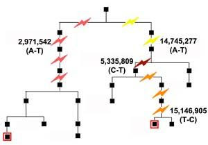
Locations of the four confirmed mutations in the family.
An international team of 16 scientists today reports the first direct measurement of the general rate of genetic mutation at individual DNA letters in humans. The team sequenced the same piece of DNA - 10,000,000 or so letters or 'nucleotides' from the Y chromosome - from two men separated by 13 generations, and counted the number of differences. Among all these nucleotides, they found only four mutations.In 1935 one of the founders of modern genetics, J. B. S. Haldane, studied men in London with the blood disease haemophilia and estimated that there would be one in 50,000 incidence of mutations causing haemophilia in the gene affected - the equivalent of a mutation rate of perhaps one in 25 million nucleotides across the genome. Others have measured rates at a few further specific genes or compared DNA from humans and chimpanzees to produce general estimates of the mutation rate expressed more directly in nucleotides of DNA.

Remarkably, the new research, recently published in Current Biology, shows that these early estimates were spot on - in total, we all carry 100-200 new mutations in our DNA. This is equivalent to one mutation in each 15 to 30 million nucleotides. Fortunately, most of these are harmless and have no apparent effect on our health or appearance.

"The amount of data we generated would have been unimaginable just a few years ago," says Dr Yali Xue from the Wellcome Trust Sanger Institute and one of the project's leaders. "But finding this tiny number of mutations was more difficult than finding an ant's egg in the emperor's rice store."

Team member Qiuju Wang recruited a family from China who had lived in the same village for centuries. The team studied two distant male-line relatives - separated by thirteen generations - whose common ancestor lived two hundred years ago.

To establish the rate of mutation, the team examined an area of the Y chromosome. The Y chromosome is unique in that, apart from rare mutations, it is passed unchanged from father to son; so mutations accumulate slowly over the generations.

Despite many generations of separation, researchers found only 12 differences among all the DNA letters examined. The two Y chromosomes were still identical at 10,149,073 of the 10,149,085 letters examined. Of the 12 differences, eight had arisen in the cell lines used for the work. Only four were true mutations that had occurred naturally through the generations.

We have known for a long time that mutations occur occasionally in each of us, but have had to guess exactly how often. Now, thanks to advances in the technology for reading DNA, this new research has been possible.

Understanding mutation rates is key to many aspects of human evolution and medical research: mutation is the ultimate source of all our genetic variation and provides a molecular clock for measuring evolutionary timescales. Mutations can also lead directly to diseases like cancer. With better measurements of mutation rates, we could improve the calibration of the evolutionary clock, or test ways to reduce mutations, for example.

Even with the latest DNA sequencing technology, the researchers had to design a special strategy to search for the vanishingly rare mutations. They used next-generation sequencing to establish the order of letters on the two Y chromosomes and then compared these to the Y chromosome reference sequence.

Having identified 23 candidate SNPs - or single letter changes in the DNA - they amplified the regions containing these candidates and checked the sequences using the standard Sanger method. A total of four naturally occurring mutations were confirmed. Knowing this number of mutations, the length of the area that they had searched and the number of generations separating the individuals, the team were able to calculate the rate of mutation.

"These four mutations gave us the exact mutation rate - one in 30 million nucleotides each generation - that we had expected," says the study's coordinator, Chris Tyler-Smith, also from The Wellcome Trust Sanger Institute. "This was reassuring because the methods we used - harnessing next-generation sequencing technology - had not previously been tested for this kind of research. New mutations are responsible for an array of genetic diseases. The ability to reliably measure rates of DNA mutation means we can begin to ask how mutation rates vary between different regions of the genome and perhaps also between different individuals."

This work was supported by the Joint Project from the NSFC and The Royal Society, and the Wellcome Trust.

Single Gene Lets Bacteria Jump From Host To Host

Share


All life — plants, animals, people — depends on peaceful coexistence with a swarm of microbial life that performs vital services from helping to convert food to energy to protection from disease.Now, with the help of a squid that uses a luminescent bacterium to create a predator-fooling light organ and a fish that uses a different strain of the same species of bacteria like a flashlight to illuminate the dark nooks of the reefs where it lives, scientists have found that gaining a single gene is enough for the microbe to switch host animals.

The finding, reported this week (Feb. 1) in the journal Nature by a team of scientists from the University of Wisconsin-Madison, is important not only because it peels back some of the mystery of how bacteria evolved to colonize different animals, but also because it reveals a genetic pressure point that could be manipulated to thwart the germs that make us sick.

"It seems that every animal we know about has microbes associated with it," says Mark J. Mandel, the lead author of the study and a postdoctoral fellow in the UW-Madison School of Medicine and Public Health. "We pick up our microbial partners from the environment and they provide us with a raft of services from helping digestion to protection from disease."

In the Pacific, a species of bacteria known as Vibrio fischeri lives in luminescent harmony with two distinct hosts: the diminutive nocturnal bobtail squid and the reef-dwelling pinecone fish. In the squid, which feeds at night near the ocean surface, one strain of the bacterium forms a light organ that mimics moonlight and acts like a cloaking device to shield the squid from hungry predators below. In the pinecone fish, another strain of the bacterium colonizes a light organ within the animal's jaw and helps illuminate the dark reefs in which it forages at night. The fish light organ may also play a role in attracting the zooplankton that make up the pinecone fish's menu.

But how did a single species of bacteria come to terms with such different hosts?

Working in the UW-Madison laboratory of microbiologist Ned Ruby, Mandel and his colleagues scoured the genomes of the two different strains of V. fischeri and found that most of the bacterium's genetic architecture was conserved over the course of millions of years of evolutionary history, but with a key difference: The strain that colonizes the squid has a regulatory gene that controls other genes that lay down a biofilm that allows the microbe to colonize the animal's light organ.

"During squid colonization, this regulatory gene turns on a suite of genes that allow bacteria to colonize the squid through mucus produced by the animal," Mandel explains. "The mucus is the pathway to the light organ, but it also helps keep out the bad guys."

Both strains of bacteria, Mandel explains, have the same genes that produce the biofilms the bacterium needs to get established in its host. But the regulatory gene that sets the other biofilm genes in motion is absent in the strain that lives in the pinecone fish, the animal scientists believe was first colonized by V. fischeri before it moved in to the squid light organ when the squid family came onto the scene in the Pacific Ocean at least 30 million years ago.

"The regulatory gene entered the bacterium's lineage and allowed it to expand its host range into the squid," according to Mandel. "The bottom-line message of the paper is that bacteria can shift host range by modifying their capabilities with small regulatory changes."

The regulatory gene acquired by the bacterium, notes Ruby, is essentially a switch the organism uses to activate a set of genes that had been residing quietly in the V. fischeri genome. Such mechanisms, he says, are very likely at play in many other species of bacteria, including those that infect humans and cause illness.

"This is going to inform a question that has been around a long time in the area of pathogenesis," says Ruby. One line of thought is that "in order to become a pathogen, a whole suite of genes needs to be imported to a bacterium."

The new finding by his group, however, suggests that nature is far more parsimonious: Instead of requiring organisms to acquire many new genes to occupy a new host, the combination of a new regulatory gene and genes that already reside in a bacterium is enough to do the trick.

"Together, they can do something neither of them could do before. They can mix and match and open up new niches," says Ruby.

Knowing that a regulatory gene plays a key role in allowing an organism to fit a new host may prove useful in human medicine as many bacterial pathogens arose first in other animals before infecting humans. A single gene can be a much easier target for a drug or other intervention to prevent or mitigate infection, the Wisconsin scientists say.

In addition to Ruby and Mandel, authors of the new Nature report include Michael S. Wollenberg, also of UW-Madison; Eric V. Stabb of the University of Georgia; and Karen L. Visick of Loyola University Chicago. The study was supported by grants from the Betty and Gordon Moore Foundation, the National Institutes of Health and the National Science Foundation.

DNA Component Can Stimulate And Suppress Immune Response

Share


A component of DNA that can both stimulate and suppress the immune system, depending on the dosage, may hold hope for treating cancer and infection, Medical College of Georgia researchers say.Low levels of CpG increase inflammation, part of the body's way of eliminating invaders. But high doses block inflammation by increasing expression of the enzyme indoleamine 2,3 dioxygenase, or IDO, an immunosuppressor, the researchers say.

"The same therapy can have two different effects," says Rusty Johnson, a fifth-year M.D./Ph.D. student in the MCG Schools of Medicine and Graduate Studies. "It was assumed that giving this treatment at higher doses would cause more stimulation, but it has the opposite effect."

The researchers hope that manipulating the dosage can help them optimize the role of inflammation in fighting invaders such as tumors and harmful bacteria. Mr. Johnson presented the findings at the Midwinter Conference of Immunologists this month in Asilomar, Calif. He is working with Drs. Andrew Mellor and David Munn, co-directors of the School of Medicine Immuno Discovery Institute, who discovered IDO's immunosuppressive capabilities more than a decade ago.

With the help of Drs. Babak Baban and Phillip Chandler, scientists in MCG's Immunotherapy Center, they've also learned IDO inhibits inflammation by blocking production of interleukin 6, a secreted factor that causes inflammation.

"This suggests that IDO is a counter-regulatory mechanism that serves as a balance to prevent too much inflammation," Mr. Johnson says. "Too much inflammation leads to destruction of normal body tissue, and this shows IDO's importance in preventing this from occurring."

The researchers already knew that IDO protects tumors from the immune system. While working with collaborators Drs. Alex Muller and George Prendergast at the Lankenau Institute in Philadelphia, they learned its role in tumor formation.

"Without it, a mouse becomes resistant to skin tumor formation, and tumors that do form are smaller and less malignant," Mr. Johnson says.

They've also learned that the cells IDO uses to suppress the immune system – IDO-competent dendritic cells – originate from B cells, which produce antibodies to fight infection.

Mr. Johnson was in his second year of medical school when he heard Dr. Munn lecture about his and Dr. Mellor's groundbreaking discovery of IDO's role in protecting a fetus from the mother's immune system. It was at that point that the Augusta native decided to pursue a career in immunology. Mr. Johnson earned a bachelor's degree in chemistry from the Georgia Institute of Technology and studied piano and voice at Augusta State University prior to coming to MCG.